Chylothorax in children and neonates: A contemporary review of controversies.

 Chyle is derived from the Greek word χυλός (chylos) which simply means juice or fluid and was used to describe digested food juices in the body by the ancient Greeks.
The clinical entity of chylothorax was first documented by a 17th century physician, anatomist and chemist, Fabrizio Bartoletti in 1633 (1) who characterised it as a ‘rare condition with a grave prognosis’ when he noticed a patient with strange, milky fluid leaking from their chest.
What Counts as Diagnostic of Chylothorax?
Milky fluid from the chest tube output or diagnostic aspiration that fulfils these biochemical criteria:
- ↑↑elevated trigylcerides (>110mg/dL or 1.24 mmol/L) in the presence of chylomicrons,
- an absolute cell count of >1000 cells/µL and a predominance of lymphocytes (>80%)
These are criteria derived from adult literature that have not been modified in over half a century and have been adopted for paediatric use. This standard biochemical cut-off becomes less reliable if the patient is fasting for prolonged periods or has never been enterally fed long chain triglycerides. This may well apply to neonates for various reasons. Although the fluid is expected to be milky-white, <50% of cases have this feature so even this may be an unreliable indicator (2,3). A fluid-to-serum cholesterol ratio of <1 and triglyceride ratio >1 is also found in chylothorax and is useful to distinguish from pseudo-chylothorax (4).
A more definitive test would be to demonstrate chylomicrons in the pleural fluid via a lipoprotein electrophoresis regardless of the infant’s feeding history (5) However, this test is expensive and may not always be readily available. An easier recommendation then would be to analyse the LDH and protein levels in the pleural fluid as most chylous effusions become protein discordant exudates. In chylothorax, protein concentration is higher than the LDH concentration (6).
Figure 2 illustrates possible causes of chylothorax in infants and children. One specific cause and scenario seen at King’s College Hospital has been the pleural effusion seen after repair of a left-sided diaphragmatic hernia. While this is common and presumably reactive in many, it can actually be a chylothorax, particularly in those coming to drainage because of pressure effects etc. (7).
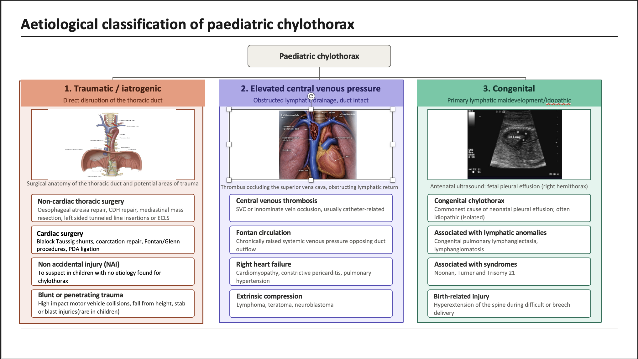
MCT Diet vs. Complete Gut Rest & Parenteral Nutrition – Which 1st-Line Management Approach to Choose?
As compared to long-chain triglycerides (LCT), medium-chain triglycerides (MCT) are transported directly into the portal circulation, does not contribute much to chylomicron formation and hence should minimise the leak.
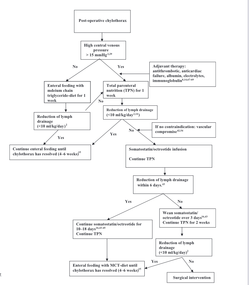
There are no RCTs to prove one is superior to another. There are a few case-control studies in post-operative chylothorax in children, however these show that there is no real difference between strategies (8–11). An MCT diet may be preferred over the side- effects that prolonged TPN can induce unless this is not an option (12).
If the chylous drainage does not decrease by the end of the first week, then converting to TPN may be a better bet. Treatment success would be characterized by decrease in the fluid effluent to <10ml/kg by the end of the first week and complete cessation by the end of second week of treatment (12). Once chylous drainage has decreased to <5mls/kg, the MCT diet can be reinstated and transition to a normal diet done over the next 2-4 weeks.
Should We Use Octreotide at All?
There is some evidence from case studies of a positive outcome from octreotide/somatostatin. However, it is difficult to say whether this was as a result of the natural history of the disease and spontaneous closure (13–15). The current level of evidence for usage of octreotide is Level 4. There has been considerable variation in the dosages used, starting from 3.5 mcg/kg/hr increased in a stepwise fashion up to 15 mcg/kg/hr for somatostatin and octreotide at 0.5 mcg/kg/hr due to its longer half-life increased to a maximum of 10mcg/kg/hr.
Neonates have been reported to develop NEC in some reports with octreotide due to splanchnic ischemia (16,17).
In conclusion, octreotide may be considered as an adjunct perhaps in the second week of conservative management of chylothorax if the output continues to remain high. However, caution needs to be exercised especially in neonates and those with pre-existing vascular disease.
When Do We Put an End to Conservative Management?
Studies have shown a success rate of about 80% with dietary modification or parenteral nutrition especially if those with elevated central venous pressure have been excluded (18, 19). Pleural fluid drainage at >10mls/kg at the end of 3-4 weeks of optimal conservative treatment should indicate failure with this method and invasive options should then be sought. Situations where there are severe metabolic or nutritional complications may necessitate earlier intervention. Patients with major vein thrombosis or high pressures in the SVC are more likely to fail conservative measures and hence this should be investigated with further imaging and echocardiography (19).
What Invasive Options are Available and Which One is Superior or Feasible?
- Pleurodesis:
Instillation of Povidone iodine (19) or fibrin glue.
- Thoracic duct embolization:
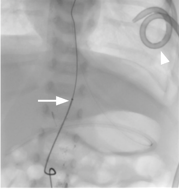
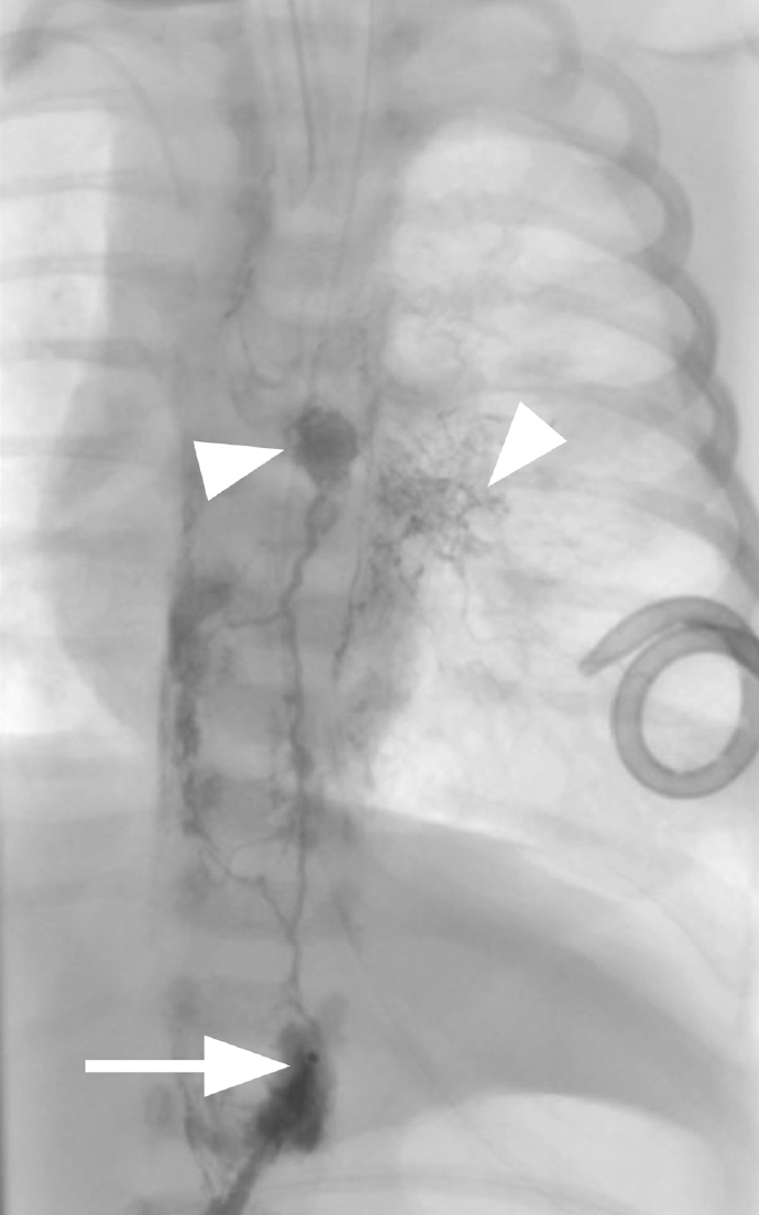
This is a minimally invasive percutaneous procedure that is an alternative to thoracic duct ligation (20). This requires intranodal lymphangiography to opacify the lymphatic system. Once the target duct is identified after injecting lipiodol through the groin with a spinal needle, an intra-abdominal cannulation to access an upper abdominal lymphatic is then performed to reach the thoracic duct and fluoroscopy images are obtained. (Figs:3,4)
Surgical
- Mechanical pleurodesis via thoracotomy or thoracoscopy
- Ligation of thoracic duct with intraoperative localisation utilising perioperative administration of cream or olive oil.
- Mass ligation of the site where the thoracic duct is presumed to insert is indicated if the duct cannot be localised.
Pleuroperitoneal shunts
These have been described since the 1980s for neonates up to 3 months of age with persistent chylothorax. So, Engum et al. from Indianapolis described placements of the Denver shunt in 7 infants with refractory chylothoraces (21).
Diaphragmatic fenestration
This unusual solution has been reported following recalcitrant chylothorax post-cardiac surgery. Kumar et al. describe 8 infants that had fenestration achieved by creation of a central diaphragmatic defect, almost invariably on the right, and suturing a Gore-Tex patch with holes deliberately cut in to the margins to restore integrity but allow fluid to leak into the peritoneal cavity (22).
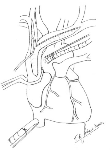
Superior Vein thrombectomy for those with SVC Outflow Tract Obstruction
These are usually post-cardiac surgery in origin. Kumar et al. described 4 cases where a thrombectomy was carried out following a long venotomy from the SVC into the brachiocephalic veins while on cardio-pulmonary by-pass, closure of the SVC achieved with a homologous patches (22). (Fig. 5)
Congenital Chylothorax
These can be isolated or associated with syndromes e.g. Trisomy 21, Turner, Noonan and monosomy X syndromes.
- Prenatal management with thoracocentesis, thoracoamniotic shunting or pleurodesis with OK-432.
- Postnatal management
- Management follows the same principles as for acquired chylothorax. Adjuncts to medical management include:
-
-
- Propranolol therapy (including antenatal therapy) may be efficacious in treating congenital chylothorax associated with lymphatic malformations by suppressing proangiogenic/lymphangiogenic factors (23).
- Sildenafil and sirolimus have been described in case reports but dosing information is limited and further evaluation is necessary before instituting these drugs (24).
-
-
Further Reading of Consensus Recommendations/ Ongoing Trials:
Trials are currently enrolling specifically within the post-cardiac surgery population except perhaps the EURO-LYMPH study and whether cardiac-surgery trial data can be extrapolated to non-cardiac traumatic or congenital chylothorax remains unclear and warrants dedicated study.
1. Development of consensus recommendations for the management of post-operative chylothorax in paediatric congenital heart disease: Quality initiative representing 22 centres in USA that describes a consensus for a management algorithm for children who develop chylothorax within 30 days of a cardiac surgery.. (https://pubmed.ncbi.nlm.nih.gov/35792060/)
2. Propranolol for postoperative chylothorax after cardiac surgery: Prospective, randomised, double blind registered clinical trial in New York on the premise that cardiac anomalies may have intrinsically abnormal lung lymphatics. Expected to complete recruitment by 2031. (https://clinicaltrials.gov/study/NCT06833320#study-overview)
3. EURO-LYMPH: Multicentre observational study that aims to evaluate the real-world use, timing, and outcomes of lymphatic interventions in patients with congenital heart disease and/or primary lymphatic disorders.Expected to end recruitment in 2030.
( https://clinicaltrials.gov/study/NCT07131293)
Summary
References
- Royal College of Physicians of Edinburgh – Education portal [Internet]. RCPE heritage -Top tips for the on call medic. Available from: https://learning.rcpe.ac.uk/mod/hvp/view.php?id=16185
- Basha AK, Prakash A, Singh KK, Prajapat D, Talwar D, Talwar D. Beyond the Milky Pleural Fluid: A 14-Year study on non-traumatic chylothorax from a tertiary care hospital in India. Cureus. 2025;17(3):e80381. doi:10.7759/cureus.
- Ruangnapa K, Anuntaseree W, Saelim K, et al. Treatment and outcomes of chylothorax in children: 20-year experience of a single institute. J Thorac Dis. 2022 Oct;14(10):3719–26. doi:10.21037/jtd-22-474
- Nair SK, Petko M, Hayward MP. Aetiology and management of chylothorax in adults. Eur J Cardio-Thorac Surg Off J Eur Assoc Cardio-Thorac Surg. 2007;32(2):362–9. doi:10.1016/j.ejcts.2007.04.024
- Maldonado F, Hawkins FJ, Daniels CE, Doerr CH, Decker PA, Ryu JH. Pleural fluid characteristics of chylothorax. Mayo Clin Proc. 2009;84(2):129–33. doi:10.4065/84.2.129
- Agrawal V, Doelken P, Sahn SA. Pleural fluid analysis in chylous pleural effusion. Chest. 2008 Jun;133(6):1436–41. doi:10.1378/chest.07-2232
- Kavvadia V, Greenough A, Davenport M, Karani J, Nicolaides KH. Chylothorax after repair of congenital diaphragmatic hernia–risk factors and morbidity. J Pediatr Surg. 1998;33(3):500-2. doi: 10.1016/s0022-3468(98)90097-5.
- Allen EM, van Heeckeren DW, Spector ML, Blumer JL. Management of nutritional and infectious complications of postoperative chylothorax in children. J Pediatr Surg. 1991;26(10):1169–74. doi:10.1016/0022-3468(91)90325-n
- Chan EH, Russell JL, Williams WG, Van Arsdell GS, Coles JG, McCrindle BW. Postoperative chylothorax after cardiothoracic surgery in children. Ann Thorac Surg. 2005;80(5):1864–70. doi:10.1016/j.athoracsur.2005.04.048
- Nguyen DM, Shum-Tim D, Dobell AR, Tchervenkov CI. The management of chylothorax/chylopericardium following pediatric cardiac surgery: a 10-year experience. J Cardi Surg. 1995 ;10(4 Pt 1):302–8. doi:10.1111/j.1540-8191.1995.tb00616.x
- Panthongviriyakul C, Bines JE. Post-operative chylothorax in children: an evidence-based management algorithm. J Paediatr Child Health. 2008;44(12):716–21. doi:10.1111/j.1440-1754.2008.01412.x
- Bellini C, Cabano R, De Angelis LC, et al. Octreotide for congenital and acquired chylothorax in newborns: A systematic review. J Paediatr Child Health. 2018;54(8):840–7. doi:10.1111/jpc.13889
- Roehr CC, Jung A, Proquitté H, et al. Somatostatin or octreotide as treatment options for chylothorax in young children: a systematic review. Intensive Care Med. 2006;32(5):650–7. doi:10.1007/s00134-006-0114-9
- Das A, Shah PS. Octreotide for the treatment of chylothorax in neonates. Cochrane Database Syst Rev. 2010 Sep 8;(9):CD006388. doi:10.1002/14651858.CD006388.pub2
- Mohseni-Bod H, Macrae D, Slavik Z. Somatostatin analog (octreotide) in management of neonatal postoperative chylothorax: is it safe? Pediatr Crit Care Med J Soc Crit Care Med World Fed Pediatr Intensive Crit Care Soc. 2004;5(4):356–7. doi:10.1097/01.pcc.0000123552.36127.22
- Chandran S, Agarwal A, Llanora GV, Chua MC. Necrotising enterocolitis in a newborn infant treated with octreotide for chylous effusion: is octreotide safe? BMJ Case Rep. 2020 Feb 11;13(2):e232062. doi:10.1136/bcr-2019-232062
- Kaewchusen P, Densupsoontorn N, Kanjanauthai S, Saengpanit P. Success rates of conservative treatment and optimal surgical timing for pediatric chylothorax. Clin Exp Pediatr. 2025;68(11):871–8. doi:10.3345/cep.2025.00598
- Beghetti M, La Scala G, Belli D, et al. Etiology and management of pediatric chylothorax. J Pediatr. 2000;136(5):653–8. doi:10.1067/mpd.2000.104287
- Scottoni F, Fusaro F, Conforti A, Morini F, Bagolan P. Pleurodesis with povidone-iodine for refractory chylothorax in newborns: Personal experience and literature review. J Pediatr Surg. 2015;50(10):1722–5. doi:10.1016/j.jpedsurg.2015.03.069
- Chick JFB, Gemmete JJ, Cline M, Srinivasa RN. Successful thoracic duct embolization for treatment of an iatrogenic left chylothorax in a neonate after repair of a tracheoesophageal fistula and esophageal atresia. J Vasc Interv Radiol. 2017;28(9):1325–7. doi:10.1016/j.jvir.2017.05.004
- Engum SA, Rescorla FJ, West KW, Scherer LR, Grosfeld JL. The use of pleuroperitoneal shunts in the management of persistent chylothorax in infants. J Pediatr Surg. 1999 ;34(2):286–90. doi:10.1016/s0022-3468(99)90192-6
- Kumar TKS, Balduf K, Boston U, Knott-Craig C. Diaphragmatic fenestration for refractory chylothorax after congenital cardiac surgery in infants. J Thorac Cardiovasc Surg. 2017;154(6):2062–8. doi:10.1016/j.jtcvs.2017.08.002
- Handal-Orefice R, Midura D, Wu JK, e at. Propranolol therapy for congenital chylothorax. Pediatrics. 2023;151(2):e2022058555. doi:10.1542/peds.2022-058555
- Hattori M, Tamai K, Watanabe H, Takeuchi A, Nakamura M, Ozeki M, Kageyama M. Sirolimus for Recurrent Chylothorax and Edema in an Infant with Noonan Syndrome after Resolved Hydrops Fetalis: A Case Report. AJP Rep. 2025 Dec 30;15(4):e186-e188. doi: 10.1055/a-2770-4952. PMID: 41476857; PMCID: PMC12753187.
Curated by Prof. Mark Davenport Sept. 2026