Persistent air leaks and bronchopleural fistulae
Ferzine Mohamed
Senior Clinical Fellow
King’s College Hospital, London
Definition
A bronchopleural fistula (BPF) is an abnormal communication between the bronchial tree and the pleural space. Though less common in the paediatric population than in adults, the consequences can be severe, and management requires a thorough understanding of the underlying aetiology, anatomy, and available therapeutic options.
The most common causes in the paediatric population are:
- Spontaneous pneumothorax,
- Empyema following a community-acquired pneumonia (1).
- Blunt or penetrating trauma.
- Post-lung surgery.
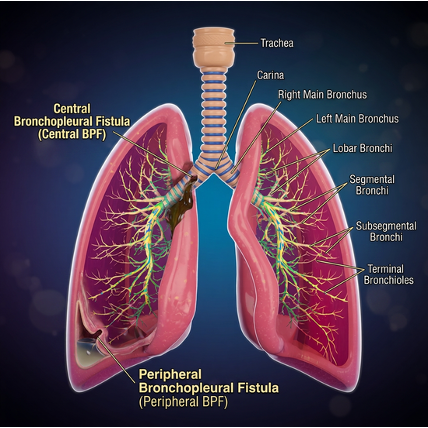
BPFs can be classified as central (mainstem, lobar, or segmental bronchus to the pleural space) or peripheral (Subsegmental bronchi, bronchioles and alveoli to the pleural space). (Fig 1)
The latter are also termed alveolopleural fistulae when the connection is alveolar rather than bronchial level. The majority of the air leaks we clinically manage are peripheral BPFs and rarely come across central BPFs. There are currently no guidelines for management of BPFs in children.
Clinical Presentation:
When a bronchopleural fistula develops, it may lead to the following:
- A persistent air leak preventing lung re-expansion ± subcutaneous emphysema
- Persistent increasing or recurrent pneumothorax despite chest drain insertion and continuous air leak through an intercostal drain (bubbling that does not resolve)
- Progressive respiratory compromise
- Risk of tension pneumothorax if the fistula acts as a one-way valve
- In ventilated patients, significant loss of tidal volume through the fistula, making ventilation extremely challenging.
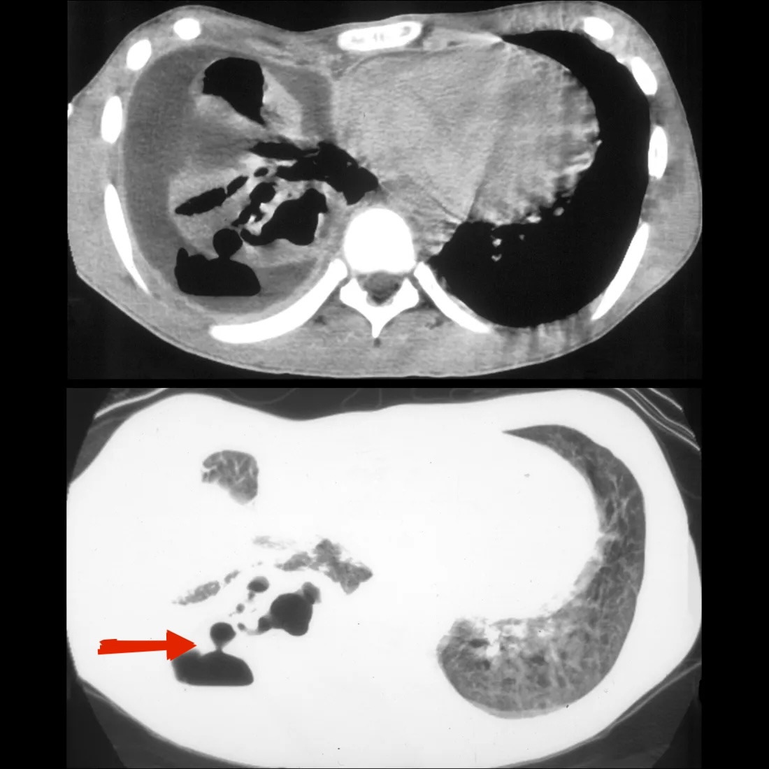
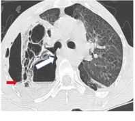
Investigations
- Chest X-ray
- always first-line investigation and it may show pneumothorax, hydropneumothorax and/or mediastinal shift but does not confirm presence and site of fistula
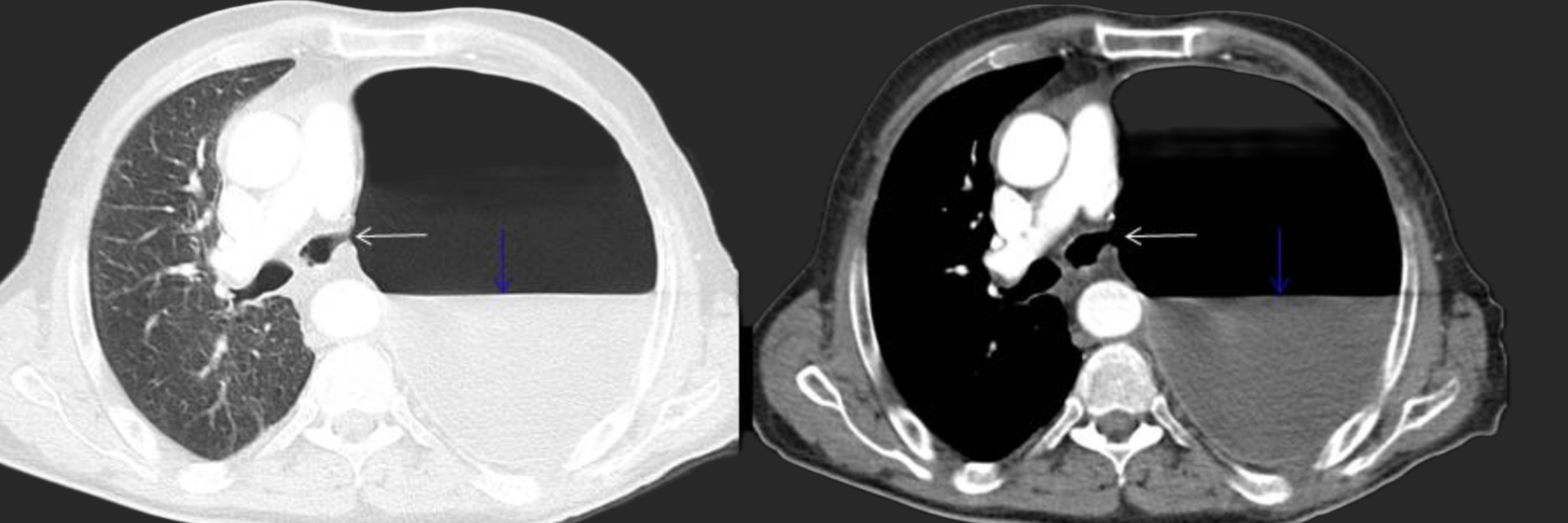
- Multidetector CT Chest (with contrast) (Fig. 4)
- Gold standard for investigation which will define anatomy, site of the fistula, assesses underlying lung parenchyma, and evaluates for collections or necrotising change.
- Gathers volumetric data in high resolution that can be used to reconstruct 3D images.
- Shows relation to adjacent vasculature, mediastinal structures, pleural space, and the diaphragm. May not always localise peripheral BPFs well. May show subtle signs of a small BPF, such as extraluminal air bubbles adjacent to the bronchial stump or a change in the appearance of a pre-existing pleural air fluid level.
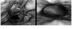
- Advanced imaging post processing applications (Fig 5)
- Virtual bronchoscopy-3D reconstruction of 2D helical CT images providing simulated endobronchial views for intraluminal evaluation of the trachea bronchial tree. Accurately shows lumen and diameter down to the 4th order bronchial branches (5,6).
- Can yield false-negative results if a fistulous tract has been plugged with mucus or debris or the images have been mis- registered during respiration.
- Ventilation scintigraphy
- Use of radioactive tracers to equilibrate in the pleural space after inhalation. However, this is time consuming and requires patient cooperation and is rarely used in children.
- Invasive tools
- Flexible fibreoptic bronchoscopy (7)
- Essential for direct visualisation of the fistula site and surrounding tissue. Useful for central BPFs. Use of a balloon occluder and pressure sensor at the tip can assess pressure and flow after the balloon has been inflated.
- Use of catheter with capnography: increase in end-tidal C02 during respiration in normal airways but flat curve in the area of the fistula.
- Flexible fibreoptic bronchoscopy (7)
Initial Management options
- Treat underlying cause with broad spectrum IV antibiotics
- Postural drainage of fluid – with the affected side on a dependent position
- Consider multiple chest drains and suction to cause apposition of pleura to chest wall.
- Application of gentle suction (8-10 cm of water)
- If on a ventilator:
- Reduce mean airway pressures, PEEP and minute ventilation.
- Lung ventilation with single lung isolation with selective bronchial intubation.
Utilisation of other techniques such differential lung ventilation, High Frequency
Management :
- Classification of air leak is useful to know severity. With conventional chest drains, the air leak is quantified by bubbling in the chest drain. A digital chest drain provides an objective measurementof air leaks and intrathoracic pressures. Table 1 illustrates a classification of severity of air leaks in clinical practice.
| Table 1: Certfolio classification of air leaks | |
| Grade 1, FE | During forced expiration only, typically when asking the patient to cough |
| Grade 2, E | Expiratory only |
| Grade 3, I | Inspiratory only |
| Grade 4, C | Continuous bubbling present in the air leak chamber during both inspiration and expiration. These leaks tend to be large and are more likely to be seen in patients receiving positive pressure ventilation |

Scenarios
- Persistent air leak for PSP (Primary spontaneous pneumothorax) after chest drain insertion but prior to surgery:
-

Fig 6: Digital suction Should suction be used to treat persistent air leak ?:
- Both the European practice and British Thoracic Society (BTS) guidelines make no recommendations for or against usage of suction in the management of spontaneous pneumothorax due to lack of strong evidence supporting one over the other. Suction levels in literature seem to be between -10 to -20 cm of H2O if being used. Jablonski et al. recommends low physiological levels of suction (-8cm H2O) (9,10). Some studies showed digital suction to be superior to wall suction.(11,12,13,14)(Fig:6)
-
- Use of blood patch/endobronchial therapies (adult practice)
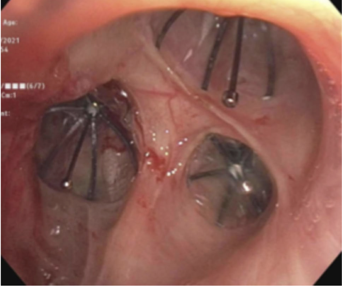
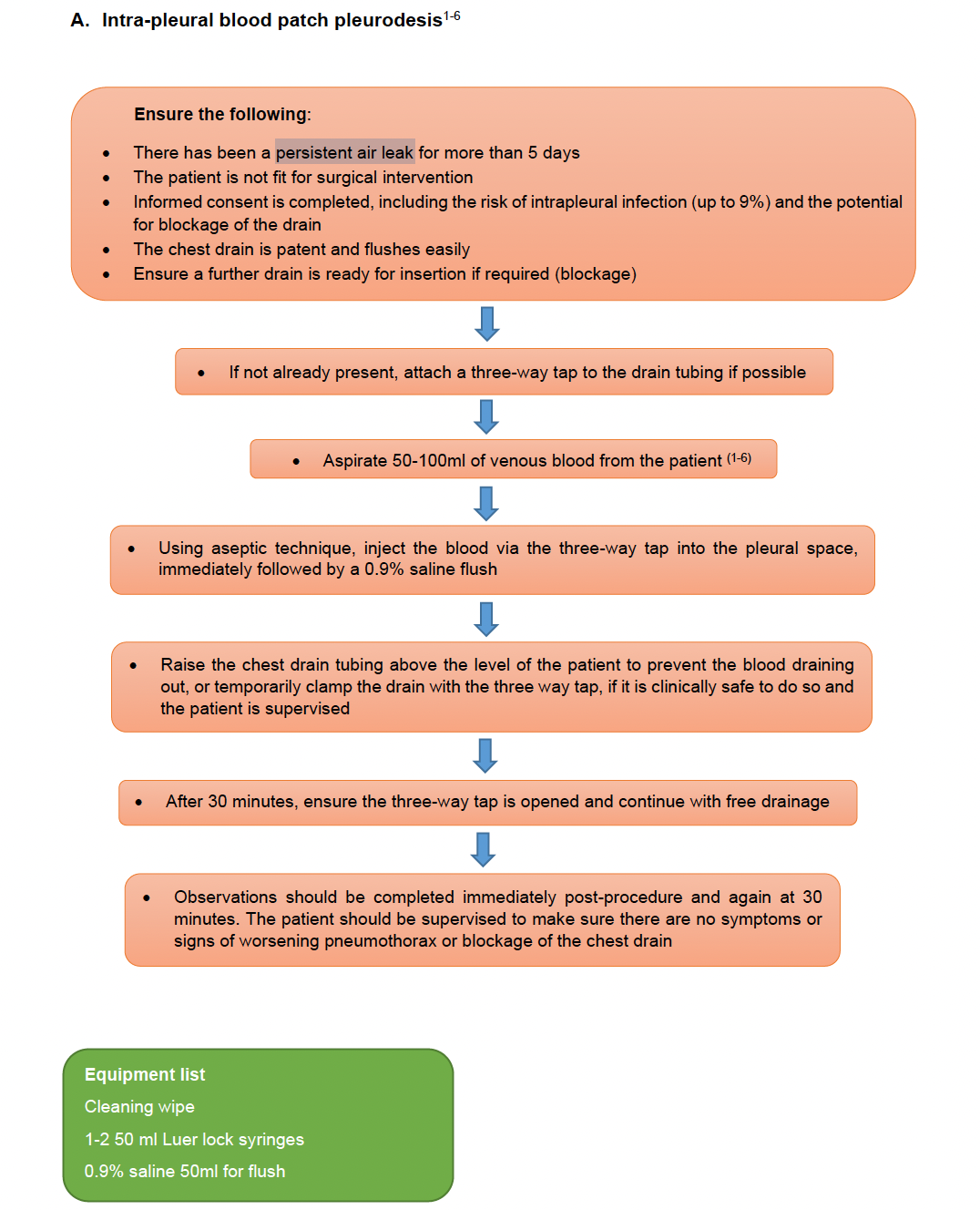
- BTS guidelines (15) recommend the use of blood pleurodesis and endobronchial therapies in patients considered unfit for surgery. ERS (16) made no recommendations for or against it. Technique of blood patch described in flowchart 1 below (BTS guidelines for pleural procedures). (17)
- Endobronchial procedures include endobronchial blood plus thrombin patch and bronchial occlusion using silicone spigots.
- Surgical options
- Resection of lung parenchyma with visible blebs or apicectomy if blebs not visible- Treatment of the underlying condition. See section on primary spontaneous pneumothorax in BAPS thoracic subspecialties.
- Surgical pleurodesis: pleural abrasion / pleural stripping / chemical pleurodesis.
- Persistent air leak after surgical procedures: excision of blebs/lobectomy/pneumonectomy: (stump leak)
- Underwater seal/suction to begin with as described above as initial options.
- Endobronchial treatment: Adult literature has described usage of Cyanoacrylate glue, sclerosing agents like silver nitrate and carbolic acid but have not been described in children.(18)

- Surgical options: Serratus Anterior, Lattisimus Dorsi muscle flaps if muscles were not divided during initial procedure. Other options are Rectus Abdominis, Pectoralis Major (great option due to dual blood supply but bulky), and intercostal muscles. The Serratus Anterior flap is probably the commonest actually used in paediatric practice (19) (Fig:8).
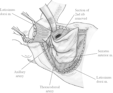
| Fig 8: Serratus anterior flap being harvested (20) |
- Persistent air leak after decortication for empyema with underlying lung parenchymal compromise:
- Can occur in up to 7.5% of procedures (21). Although high-quality randomised trial data are limited, numerous case series and observational studies support the efficacy and safety of VAC therapy in thoracic infections.
- Neonates and infants
- Options to be considered and tailored to specific patient.
- Consider any of the Initial measures as mentioned above in the box. Single lung ventilation techniques may not be possible due to the weight and gestational age of the patient.
- Pleurodesis: with fibrin glue
- Surgical options:
- Airway repair via thoracotomy (22).
- Oversewing Lobectomy
| Flowchart 1: depicting technique of blood patch pleurodesis |
REFERENCES
- McKee AJ, Ives A, Balfour-Lynn IM. Increased incidence of bronchopulmonary fistulas complicating pediatric pneumonia. Pediatr Pulmonol. 2011 Jul;46(7):717-21. doi: 10.1002/ppul.21396.
- CT Fig 2: Pediatric Imaging. Pediatric bronchopleural fistula [Internet]. 2019 Jun 20 [updated 2025 Mar 7; cited 2026 Aug 24]. Available from: https://pediatricimaging.org/diseases/bronchopleural-fistula/
- CT Fig 3: Wang XM, Guo LC, Xue SL, Chen YB. Pulmonary mucormycosis: A case report and review of the literature. Oncol Lett. 2016 May;11(5):3049-3053. doi: 10.3892/ol.2016.4370. Epub 2016 Mar 22. PMID: 27123061; PMCID: PMC4841004.
- CT Fig 4: Courtesy: Bronchopleural fistulas (BPF). ‘Radiologic information from the beginning to the end’: poster presented in Spanish at the 2010 Congreso Nacional SERAM (A Coruña)
- CT Fig 5: Miranti PI, Lubis MRS. Role of computed tomography virtual bronchoscopy in a case of pulmonary tuberculosis complicated by bronchopleural fistula and pyopneumothorax. Radiol Case Rep. 2026 May 14;21(8):3244-3248. doi: 10.1016/j.radcr.2026.04.019.
- Gaur P, Dunne R, Colson YL, Gill RR. Bronchopleural fistula and the role of contemporary imaging. J Thorac Cardiovasc Surg. 2014 Jul;148(1):341-7. doi: 10.1016/j.jtcvs.2013.11.009.
- Felipe A, Macarena R. Vial. Overview of Bronchopleural Fistula Management, with a Focus on Bronchoscopic Treatment. Curr Pulmonol Rep. 2022 Apr;11(1). doi: 10.1007/s13665-022-00289-3.
- Dugan KC, Laxmanan B, Murgu S, Hogarth DK. Management of Persistent Air Leaks. Chest. 2017 Aug;152(2):417-423. doi: 10.1016/j.chest.2017.02.020.
- George RS, Papagiannopoulos K. Advances in chest drain management in thoracic disease. J Thorac Dis. 2016 Feb;8(Suppl 1):S55-64. doi: 10.3978/j.issn.2072-1439.2015.11.19.
- Jablonski S, Brocki M, Wawrzycki M, Smigielski JA, Kozakiewicz M. Efficacy assessment of the drainage with permanent airflow measurement in the treatment of pneumothorax with air leak. Thorac Cardiovasc Surg. 2014 Sep;62(6):509-15. doi: 10.1055/s-0033-1359714.
- Walker S, Hallifax R, Ricciardi S, Fitzgerald D, Keijzers M, Lauk O, et al. Joint ERS/EACTS/ESTS clinical practice guidelines on adults with spontaneous pneumothorax. Eur Respir J. 2024 May 28;63(5):2300797. doi: 10.1183/13993003.00797-2023.
- Leo F, Duranti L, Girelli L, Furia S, Billè A, Garofalo G, Scanagatta P, Giovannetti R, Pastorino U. Does external pleural suction reduce prolonged air leak after lung resection? Results from the AirINTrial after 500 randomized cases. Ann Thorac Surg. 2013 Oct;96(4):1234-1239. doi: 10.1016/j.athoracsur.2013.04.079.
- Cerfolio RJ, Bass C, Katholi CR. Prospective randomized trial compares suction versus water seal for air leaks. Ann Thorac Surg. 2001 May;71(5):1613-7. doi: 10.1016/s0003-4975(01)02474-2.
- Lim E, Blackmon SH, Depypere L, Gonzalez M, Mitzman B, Stiles BM, Novoa N. Clinical outcomes of digital versus traditional chest drains after thoracic surgery: a narrative review of randomised trials. J Thorac Dis. 2025 Aug 31;17(8):6318-6325. doi: 10.21037/jtd-2025-298.
- BTS guidelines: Online appendix 12 (Pleural procedures)_intrapleural treatment.pdf
- Walker S, Hallifax R, Ricciardi S, et al. Joint ERS/EACTS/ESTS clinical practice guidelines on adults with spontaneous pneumothorax. European Respiratory Journal. Published by the European Respiratory Society (ERS), European Association for Cardio-Thoracic Surgery (EACTS), and European Society of Thoracic Surgeons (ESTS), 2024.
- BTS guidelines: Online appendix A3 (Pleural disease)_Pneumothorax ongoing air leak management
- Cerfolio RJ. Advances in thoracostomy tube management. Surg Clin North Am. 2002 Aug;82(4):833-48, vii. doi: 10.1016/s0039-6109(02)00026-9. PMID: 12472132.
- Jester I, Nijran A, Singh M, Parikh DH. Surgical management of bronchopleural fistula in pediatric empyema and necrotizing pneumonia: efficacy of the serratus anterior muscle digitation flap. J Pediatr Surg. 2012 Jul;47(7):1358-62. doi: 10.1016/j.jpedsurg.2011.12.012.
- Fig 8: Groth SS, Whitson BA, D’Cunha J, Andrade RS, Landis GH, Maddaus MA. Serratus anterior transposition muscle flaps for bronchial coverage: technique and functional outcomes. Ann Thorac Surg. 2009 Dec;88(6):2044-6. doi: 10.1016/j.athoracsur.2009.04.062.
- Scheese D, Freudenberger DC, Mastoloni E, et al. Reducing air leak after empyema surgery: COPD’s role and patient management. J Surg Res 2025; 307: 116–121. doi:1016/j.jss.2025.01.020
- Milan A, Yardley I, Fox G, Meau-Petit V. Stepwise multidisciplinary approach in very low birthweight infant with bronchopleural fistula. BMJ Case Rep. 2021 Jul 12;14(7):e241721. doi: 10.1136/bcr-2021-241721.
Curated by Prof. Mark Davenport
September 2026